Digital X-Ray
Naperville, IL
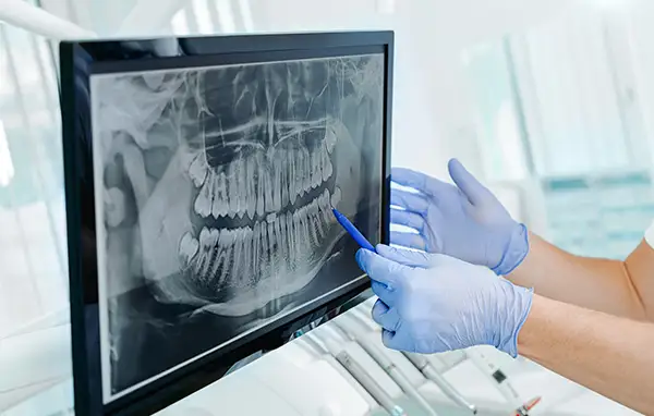 When a dentist takes a digital X-ray, the process looks simple, a small sensor is placed in the mouth, a button is pressed, and an image appears on screen within seconds. Behind that simplicity is engineering that has quietly transformed dental diagnostics. Understanding what's happening inside that sensor, and how it differs from the film-based systems dentistry relied on for most of the last century, helps explain why digital imaging has become the standard of care almost everywhere. When a dentist takes a digital X-ray, the process looks simple, a small sensor is placed in the mouth, a button is pressed, and an image appears on screen within seconds. Behind that simplicity is engineering that has quietly transformed dental diagnostics. Understanding what's happening inside that sensor, and how it differs from the film-based systems dentistry relied on for most of the last century, helps explain why digital imaging has become the standard of care almost everywhere.
For decades, dental offices depended on photographic film that had to be exposed, removed from the mouth, and carried to a darkroom for chemical development. The process took several minutes per image, tied up staff time, and produced a physical piece of film that could fade, tear, or get misplaced over the years. Digital sensors replaced that entire workflow with an electronic process that captures, processes, and displays an image before you've even had time to sit back in the chair.
Two Main Types of Digital Sensors
Dental practices generally rely on one of two sensor technologies. The first is a direct digital sensor, built with either charge-coupled device, or CCD, technology or complementary metal-oxide-semiconductor, or CMOS, technology. These sensors are wired directly to a computer and produce an image the moment the exposure is taken, converting X-ray energy into an electronic signal that a computer translates instantly into a viewable image. Because the sensor is tethered by a cable, the image appears on screen without any additional handling, which makes this option popular in practices that prioritize speed and a streamlined workflow.
CCD and CMOS sensors differ slightly in how they convert light and X-ray energy into electronic signals, but both are considered direct digital systems because no intermediate scanning step is required. CMOS sensors have become increasingly common in newer equipment because they tend to be more energy efficient and often allow for a thinner sensor design, which can improve patient comfort during placement.
The second type is a phosphor storage plate, an indirect digital system that temporarily stores X-ray energy after exposure, then is fed into a scanning device that reads the stored energy and converts it into a digital image within moments. Phosphor plates tend to be thinner and more flexible than wired sensors, which can make them more comfortable for patients with smaller mouths, a strong gag reflex, or dental anxiety related to intraoral equipment. Because they aren't attached to a cable, they also allow for slightly more flexible positioning in certain areas of the mouth that are harder to reach.
Both systems produce a detailed digital image without film or chemical processing, but they differ in comfort, speed, and workflow. A wired sensor delivers an image almost instantly, while a phosphor plate requires an extra step of being scanned, though that step typically takes only a few seconds. Many practices use a combination of both technologies depending on the specific image being captured and the needs of the individual patient.
From Exposure to On-Screen Image
Once the sensor or plate captures the X-ray energy, software takes over, processing the raw data into a grayscale image representing the varying densities of teeth, bone, and soft tissue. Dense structures, such as enamel and bone, appear lighter, while less dense areas, such as decay or pulp tissue, appear darker. This grayscale mapping is what allows a trained eye to distinguish between healthy tooth structure and areas that may require treatment.
This is where digital imaging offers a meaningful advantage over film. Because the image exists as data rather than a fixed photograph, Dr. Scherercan adjust brightness and contrast after the fact, zoom into specific areas without losing clarity, and apply filters that highlight particular structures. A subtle area of bone loss or an early cavity that's hard to see on a standard view can often be brought into sharper focus with these tools. Some software can also apply color mapping or edge enhancement, which some dentists find helpful for identifying the boundaries of a cavity or the outline of a root canal.
The processed image is stored digitally as part of your electronic dental record, which means it never fades, tears, or gets lost in a filing cabinet the way film sometimes did. It can be pulled up instantly at any future visit for direct comparison against new images, making it easier to track subtle changes in your oral health over months or years.
Why Resolution and Clarity Matter
Image resolution refers to how much fine detail a sensor can capture, and it plays a direct role in diagnostic accuracy. Higher resolution sensors can reveal the earliest stages of decay, fine cracks in tooth structure, or subtle changes in bone density that might otherwise go unnoticed, part of why digital X-rays are so valuable for catching problems while they're still small and easier to treat. Resolution is typically measured in line pairs per millimeter, a technical specification that essentially describes how finely detailed an image can be before it starts to blur.
 Clarity also supports better communication between you and the dentist. Because the image can be displayed on a large monitor and magnified in real time, the dentist can point directly to areas of concern and walk you through the findings rather than describing them from a small piece of film. This visual conversation often helps patients better understand their treatment recommendations, since seeing the issue firsthand tends to be more convincing than a verbal description alone. Clarity also supports better communication between you and the dentist. Because the image can be displayed on a large monitor and magnified in real time, the dentist can point directly to areas of concern and walk you through the findings rather than describing them from a small piece of film. This visual conversation often helps patients better understand their treatment recommendations, since seeing the issue firsthand tends to be more convincing than a verbal description alone.
Beyond individual images, resolution and clarity also matter when comparing X-rays taken months or years apart. A high-quality image today makes it easier for the dentist to notice small changes at your next visit, whether that's the slow progression of bone loss around a tooth or the gradual development of a cavity that wasn't visible previously. This kind of long-term comparison is one of the most practical benefits of high-resolution digital imaging.
A Foundation for Accurate Diagnosis
The technology inside a digital X-ray sensor may not be something patients think about during an appointment, but it plays a direct role in the quality of care you receive. Faster processing, adjustable imaging, and higher resolution all work together to give your dentist a clearer picture of what's happening beneath the surface. If you have questions about the imaging technology used during your visit, our team at Wheaton Oral Surgery is happy to walk you through the process. Call us at (331) 204-0314 to schedule your next appointment. |


